Arrector Pili Pictures, Images and Stock Photos
Browse 50+ arrector pili stock photos and images available, or start a new search to explore more stock photos and images.
Most popular
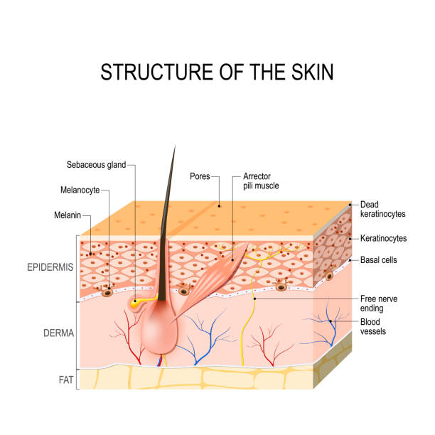
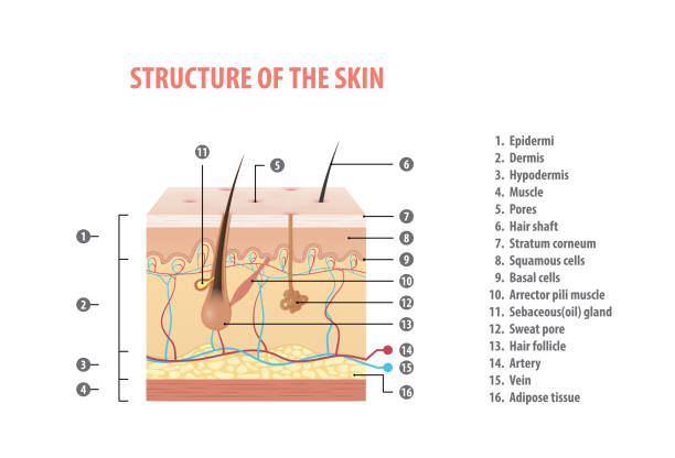
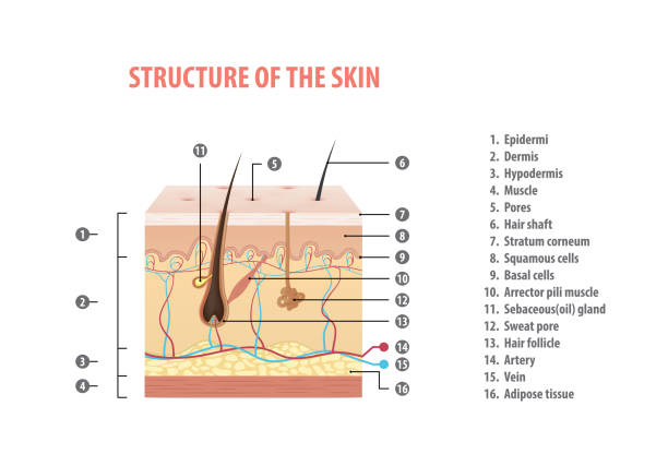
Healthy Human Skin. hair follicle, cell structure and layers. Vector illustration for your design and medical use. human anatomy.

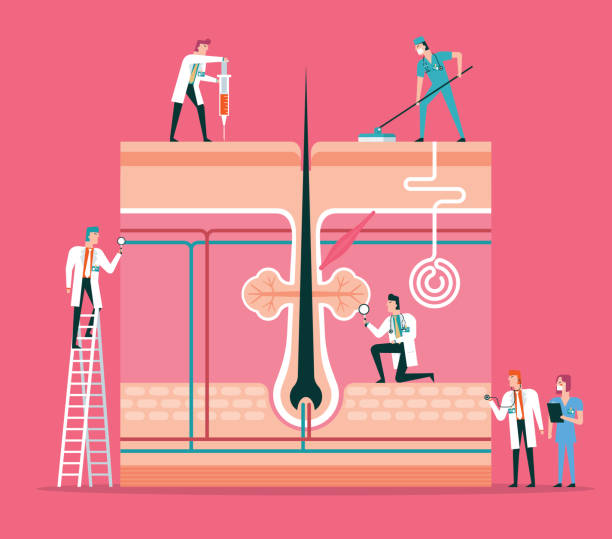
Team of doctors diagnose human skin

Team of doctors diagnose human skin
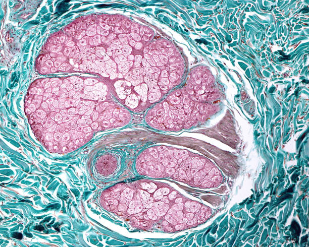
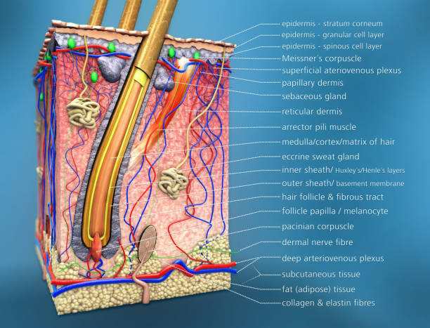
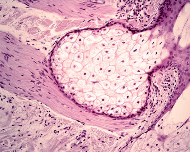
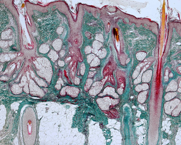
Cross section showing all the components of pilosebaceous unit: in the center, the hair, surrounded by sebaceous glands and smooth muscle fibers of arrector pili muscle. Masson stain.
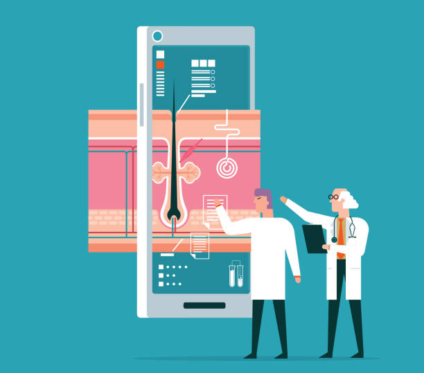
Team of doctors diagnose human skin
The sebaceous gland is a holocrine gland whose cells accumulate sebum in clear oil or lipid droplets. Finally, the cells die, which is indicated by their pyknotic nuclei, visible on top half of the image.
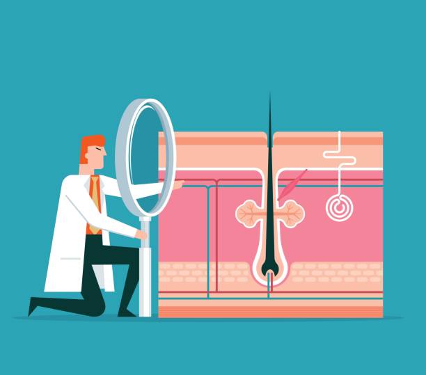
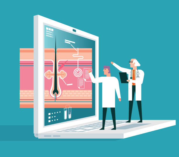
Healthcare vector concept stock illustration
Histology image of hair bulb in the dermis of skin (100x)

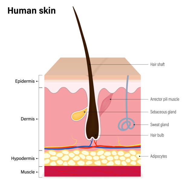

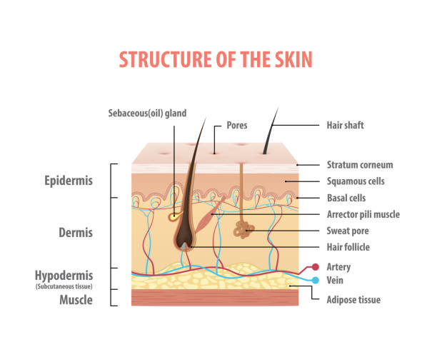
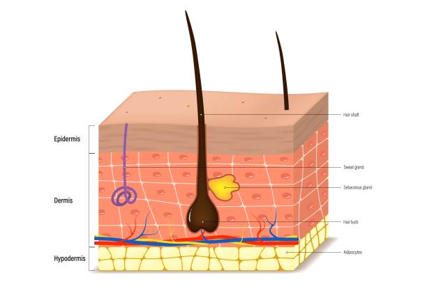
Illustration of the anatomy of human skin and hair

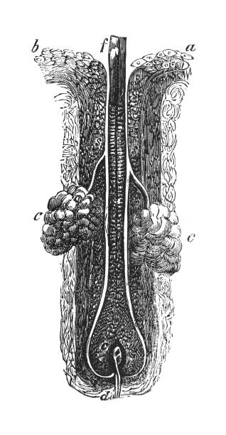
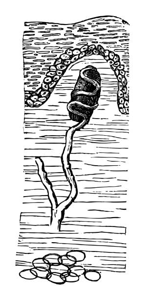
Vintage engraved illustration isolated on white background - Anatomical structure of the human hair (intersection)
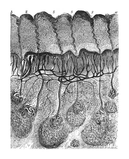
Vintage engraved illustration - Layers of the human skin (intersection)

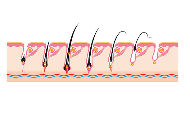
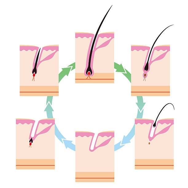
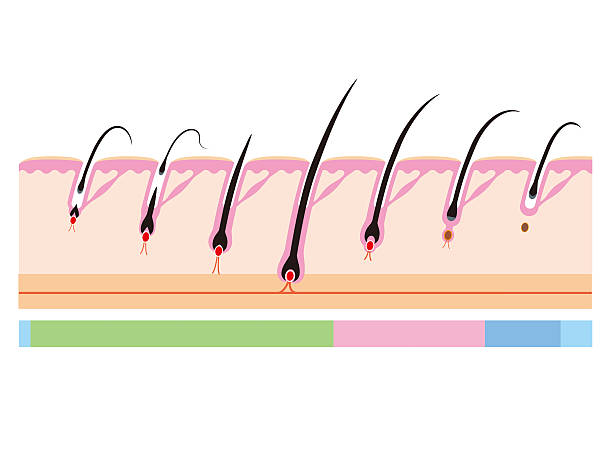
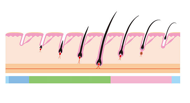
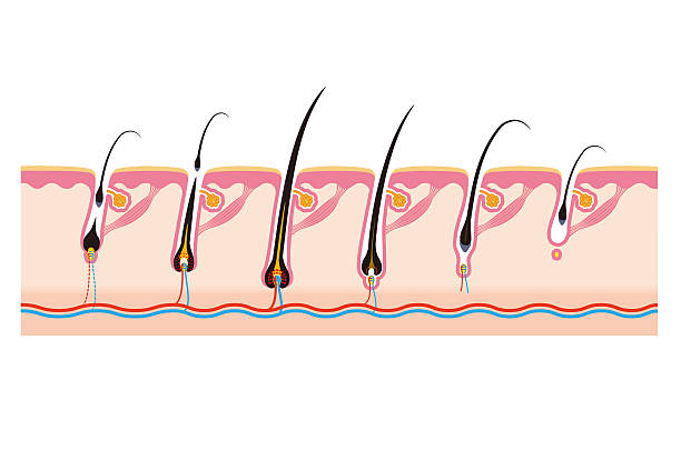
Hair growth process stages with anatomical phases structure outline diagram. Labeled educational early anagen, catagen or telogen cycle description vector illustration. Medical scalp surface side view
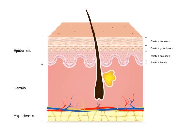
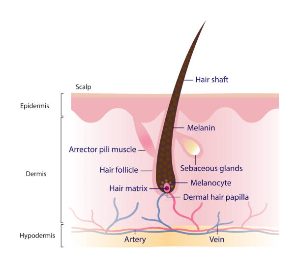
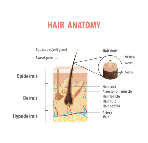
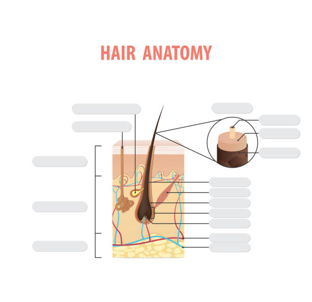
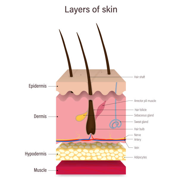
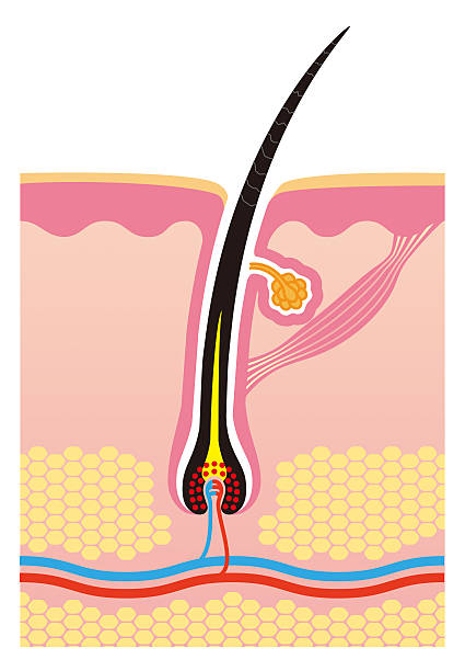
Diagram of hair structure anatomy vector illustration isolated on white background. Cross section of human hair with scalp layer. Hair shaft, arrector pili muscle, follicle, hair matrix and papilla. Skin and hair anatomy.
Vintage engraved illustration - Human skin (Tactile corpuscles or Meissner's corpuscles)
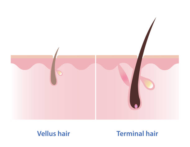
The difference between vellus hair and terminal hair vector illustration isolated on white background. Hair Types. Vellus hair is fine, wispy and unpigmented hair. Terminal hair is thick, coarse, long and pigmented hair.

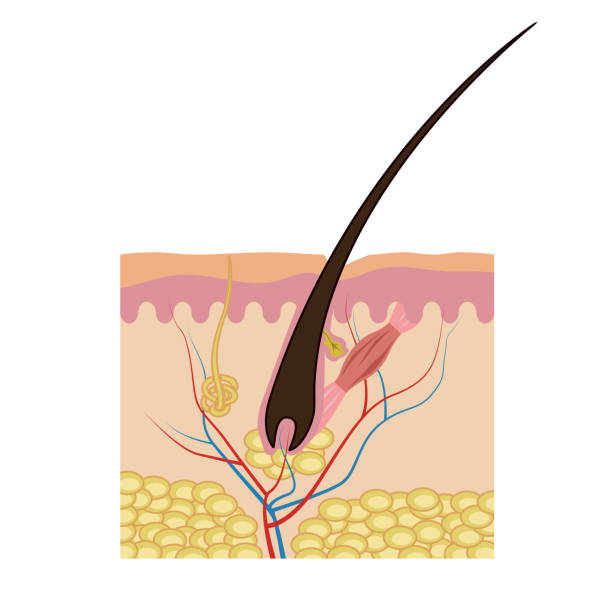
The arrector pili muscles are small bundles of smooth muscle fibres attached to hair follicles, which causes the raising of hairs known as goose bumps."n
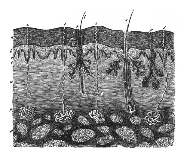
Vintage engraved illustration - Layers of the human skin


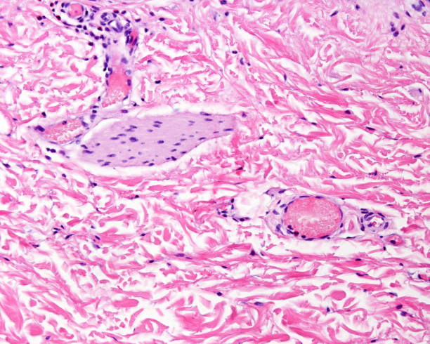
Dermis of the thin skin, rich in intermingled collagen bundles, where in can identified small blood vessels and an oblique section of the arrector pili muscle.

The sebaceous gland. It is a holocrine gland whose cells accumulate sebum in clear oil or lipid droplets. Finally, the cells die, which is indicated by their pyknotic nuclei, visible on top half of the image.
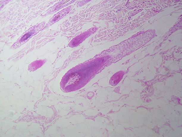
Section of scalp hairy skin showing several hair follicles with their associated sebaceous glands (pilosebaceous units) located in the dermis. Low magnification micrograph. Masson stain.
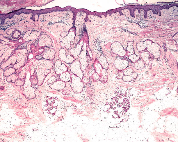
Low magnification micrograph showing the different location of the sebaceous glands (more superficial, in reticular dermis) and sweat glands (more deep, in the adipose tissue of hypodermis).
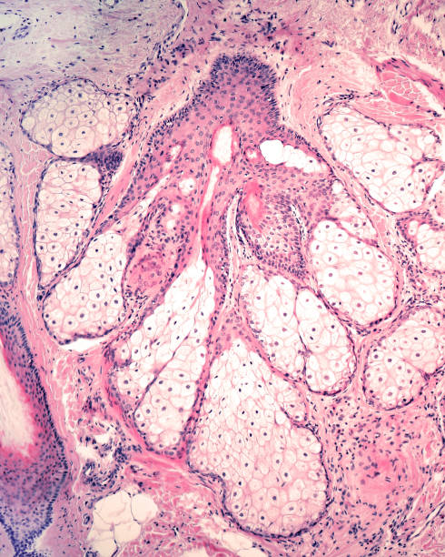
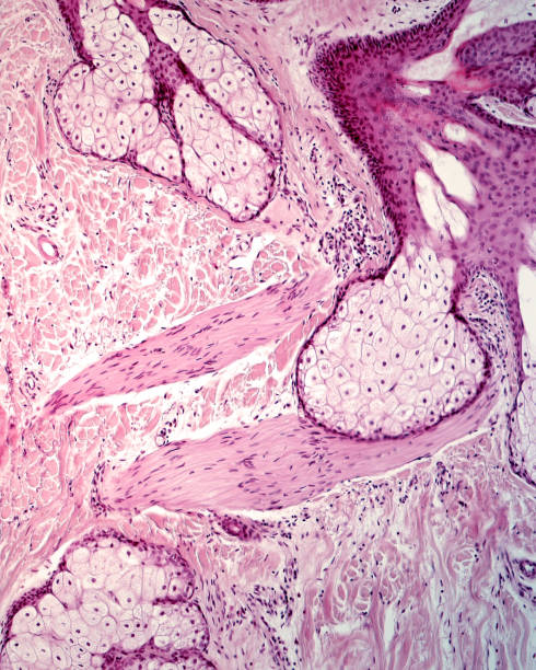
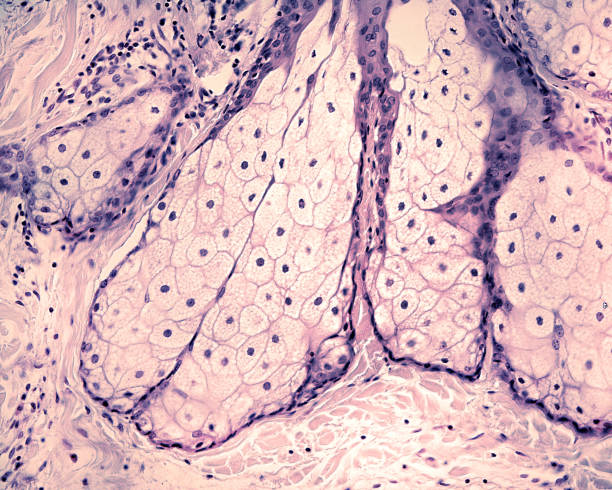
Sebaceous gland formed by multiple acinar glands branch off a central duct. In the top and bottom borders, sections of arrector pili muscles can be seen.

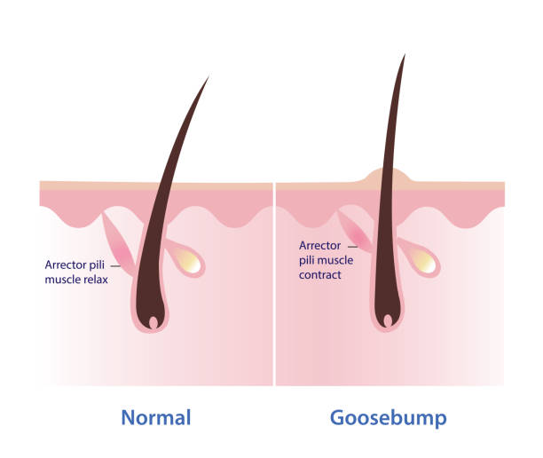
Goosebumps on the skin vector illustration isolated on white background. Comparison of normal skin, arrector pili muscle relax and goosebumps, arrector pili muscle contract and pull the hair straight up.